


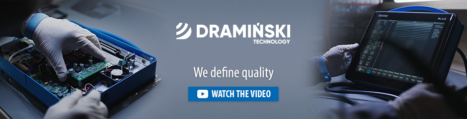


ULTRASOUND SCANNER BLUE
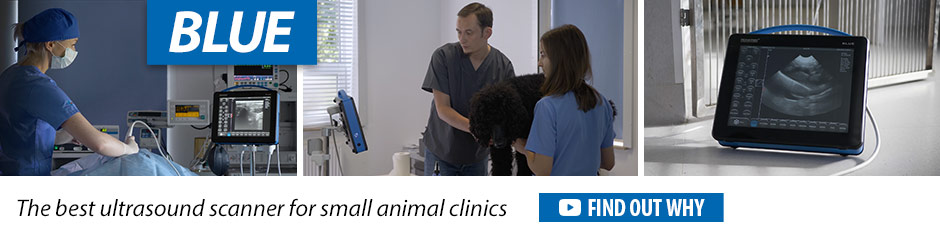
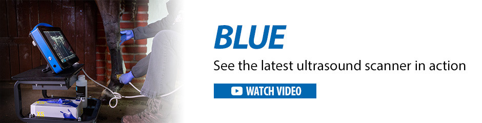
Modern ultrasound scanner with excellent Doppler ultrasonography and high quality imaging. Designed to diagnose small animals and horses.


Excellent Doppler – more precise examination
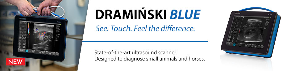
See. Touch. Feel the difference.
DRAMIŃSKI BLUE is a veterinary diagnostic ultrasound scanner that provides surprisingly detailed images. Enjoy the sensitive touch screen. The scanner is second to none after the first examination.
Technologies of the future
 |
LuciD™a system that enhances contrast, sharpness and tissue differentiation making images readable and easy to interpret. |
 |
D-Curve™distinguishes between slight echogenicity differences – a transfer curve ideally suited to the human eye perception. |
 |
Vi-Probe™is a unique technology that provides phased array image type on the convex probe and convex image on the linear probe. BLUE provides enhanced vision, even through a small acoustic window. |
BLUE Veterinary Ultrasound Scanner Features
 |
STANDBY AND OPERATING TIMEThe veterinary ultrasound scanner is ready to operate after 25 seconds. The built-in battery provides up to 2.5-h convenient operation with no power supply connection needed. |
 |
OPERATION IS SIMPLER THAN EVERActive fields allow quick standard parameter setting. Measure the distance immediately after freezing the image – no additional menu needed. Entering a patient’s data before an ultrasound examination is not necessary. This can be done while saving the first image. |
 |
3 DOPPLER IMAGING MODESThe veterinary scanner has three Doppler imaging modes: Color Doppler, Power Doppler, Pulse Wave Doppler. The technology allows examining blood flow in blood vessels. |
 |
REPLACEABLE PROBE SYSTEMIf the probe needs to be replaced during an ultrasound examination, the system will detect the new one automatically. Probes available:
|
 |
MULTITASKINGWhether it is a quick emergency screening of a patient or an accurate ultrasound diagnostics in orthopaedic surgery, BLUE will always be there, ready for new challenges. |
 |
LIGHT DESIGNLet other compromise. We have applied solutions that allow using the latest technology in the veterinary ultrasound scanner weighing 4 kg only. |
 |
LARGE TOUCH SCREENThe 12″ LED LCD display guarantees a successful diagnosis. The features are controlled via a sensitive touch panel. Quick and easy operation. |
 |
WARRANTYThe DRAMIŃSKI BLUE veterinary ultrasound scanner has a two-year warranty. |
The kit includes:
- Ultrasound scanner with built-in battery;
- Probe (optional);
- Stand;
- Power supply;
- User manual with warranty card;
- Carrying case.
What next?
Contact our account manager now.
Technical data |
||
| Imaging modes | B Mode, B+B Mode, 4B Mode, B+M Mode, Color Doppler, Power Doppler, Pulse Wave Doppler |
|
| Operating frequency | 1-14 MHz (probe type dependant) | |
| Grayscale | 256 degrees, D-Curve™ (grayscale curve adapted to the human eye perception) | |
| Image analysis and processing | LuciD™ (contrast, sharpness and tissue differentiation enhancement system) | |
| Ultrasound control |
Vi-Probe™ virtual convex probe on linear probes, virtual sector probe on convex probes | |
| Frame memory and cine loop viewing | 30 GB | |
| Data record | Images and cine loop viewing with description, patient data and date | |
| Data transmission to a data storage device | Via USB interface | |
| Exported file format | PNG for images, AVI, MP4, MOV for cine loop viewing | |
| Number of probe ports | One port, automatic probe recognition | |
| External ports | 2 x USB, 2 x LAN, 1 x HDMI | |
| Monitor | 12″ LED LCD display | |
| Feature control | Capacitive touch panel | |
| Standby time | approx. 25 s | |
| Continuous operation time with battery supply | approx. 2 hours 30 min. | |
| Package charging time | approx. 4 hours. | |
| Optional accessories | Wheel stand Caster stand (low) Gel spacer |
|
| Operating temperature | From -15°C to +45°C | |
| Recommended storage temperature | From 0°C to + 45°C | |


The Blue ultrasound offers a great image quality for OPU in horses. It also has a line that can be programmed to see where the needles goes into the ovary and the track it follows, helping a lot during the procedure. The ultrasound is easy to use, has a very light weight and has a battery that lasts for 2.5 hours. The OPU handle is also very convenient and its design helps a lot during the procedure. – Dr Macias Garcia, UEX Spain