

ULTRASOUND SCANNER BLUE
A reliable premium class portable ultrasound scanner ideal for mobile doctors.


The device is quality certified ISO 13485:2016 and EC 93/42/EEC.
Excellent Doppler – more precise examination
Quickly. Easily. Precisely.
DRAMIŃSKI BLUE is a state-of-the-art ultrasound scanner designed for doctors on call. High quality imaging, intuitive operation and light weight of the device will make it easy to take it with you to a patient. After the first try you will not return to another device.
Technologies of the future
 |
D-Curve™D-Curve™ distinguishes between slight echogenicity differences – a transfer curve ideally suited to the human eye perception. |
 |
LuciD™LuciD™ – a system that enhances contrast, sharpness and tissue differentiation making images readable and easy to interpret. |
 |
Vi-Probe™Vi-Probe™ is a unique technology that provides phased array image type on the convex probe and convex image on the linear probe. BLUE provides enhanced vision, even through a small acoustic window. |
BLUE Ultrasound Scanner Features
 |
STANDBY AND OPERATING TIMEAfter 25 seconds, the mobile ultrasound scanner is ready for operation. Built-in battery allows for 2.5 hours of continuous work. |
 |
OPERATION IS SIMPLER THAN EVERActive fields allow quick standard parameter setting. Measure the distance immediately after freezing the image – no additional menu needed. You don’t need to remember about entering a patient’s data before an ultrasound examination. This can be done while saving the first image. |
 |
3 DOPPLER IMAGING MODESThe portable scanner DRAMIŃSKI BLUE has three Doppler imaging modes: Color Doppler, Power Doppler, Pulse Wave Doppler. The technology allows examining blood flow in blood vessels. |
 |
REPLACEABLE PROBE SYSTEMIf the probe needs to be replaced during an ultrasound examination, the system will detect the new one automatically. Probes available:
|
 |
MULTITASKINGWhether it is a quick emergency screening of a patient or an accurate ultrasound diagnostics in orthopaedic surgery, BLUE will always be there, ready for new challenges. |
 |
LIGHT DESIGNLet other compromise. We have applied solutions that allow using the latest technology in this premium ultrasound scanner weighing 4 kg only. |
 |
LARGE TOUCH SCREENThe 12″ LED LCD display guarantees a successful diagnosis. The features are controlled via a sensitive touch panel. Quick and easy operation. |
 |
WARRANTYThe DRAMIŃSKI BLUE medical ultrasound scanner has a two-year warranty. |
The kit includes:
- Ultrasound scanner with built-in battery;
- Probe (optional);
- Stand;
- Power supply;
- User manual with warranty card;
- Carrying case.
What next?
Contact our account manager now.
Technical data |
||
| Dimensions | 31,0 x 28,0 x 6,5 cm | |
| Imaging modes | B Mode, B+B Mode, 4B Mode, B+M Mode, Color Doppler, Power Doppler, Pulse Wave Doppler |
|
| Operating frequency | 1-14 MHz (probe type dependant) | |
| Grayscale | 256 degrees, D-Curve™ (grayscale curve adapted to the human eye perception) | |
| Image analysis and processing | LuciD™ (contrast, sharpness and tissue differentiation enhancement system) | |
| Ultrasound control |
Vi-Probe™ virtual convex probe on linear probes, virtual sector probe on convex probes | |
| Frame memory and cine loop viewing | 30 GB | |
| Data record | Images and cine loop viewing with description, patient data and date | |
| Data transmission to a data storage device | Via USB interface | |
| Exported file format | PNG for images, AVI, MP4, MOV for cine loop viewing | |
| Number of probe ports | One port, automatic probe recognition | |
| External ports | 2 x USB, 2 x LAN, 1 x HDMI | |
| Monitor | 12″ LED LCD display | |
| Feature control | Capacitive touch panel | |
| Standby time | approx. 25 s | |
| Continuous operation time with battery supply | approx. 2 hours 30 min. | |
| Package charging time | approx. 4 hours. | |
| Optional accessories | Wheel stand Caster stand (low) Gel spacer |
|
| Operating temperature | From -15°C to +45°C | |
| Recommended storage temperature | From 0°C to + 45°C | |
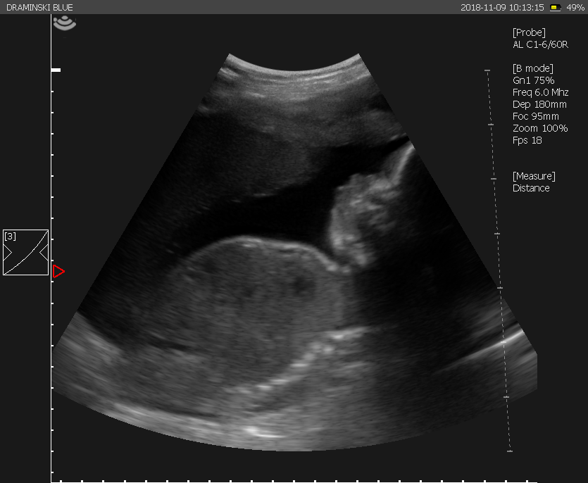
Pregnancy 31. week |
 |
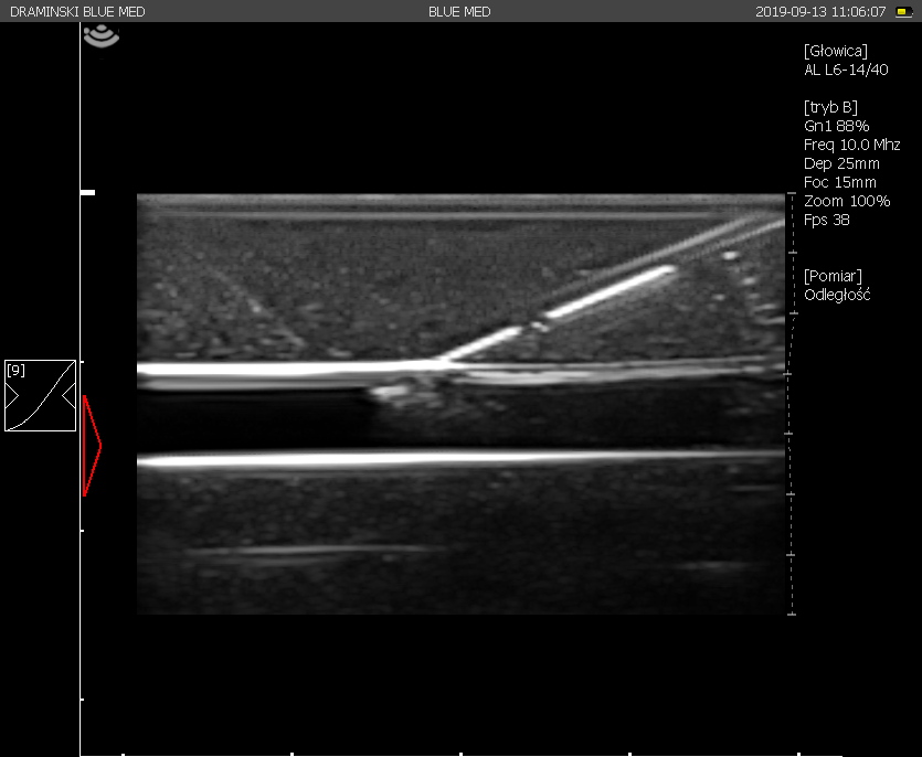
Needle insertion |
 |
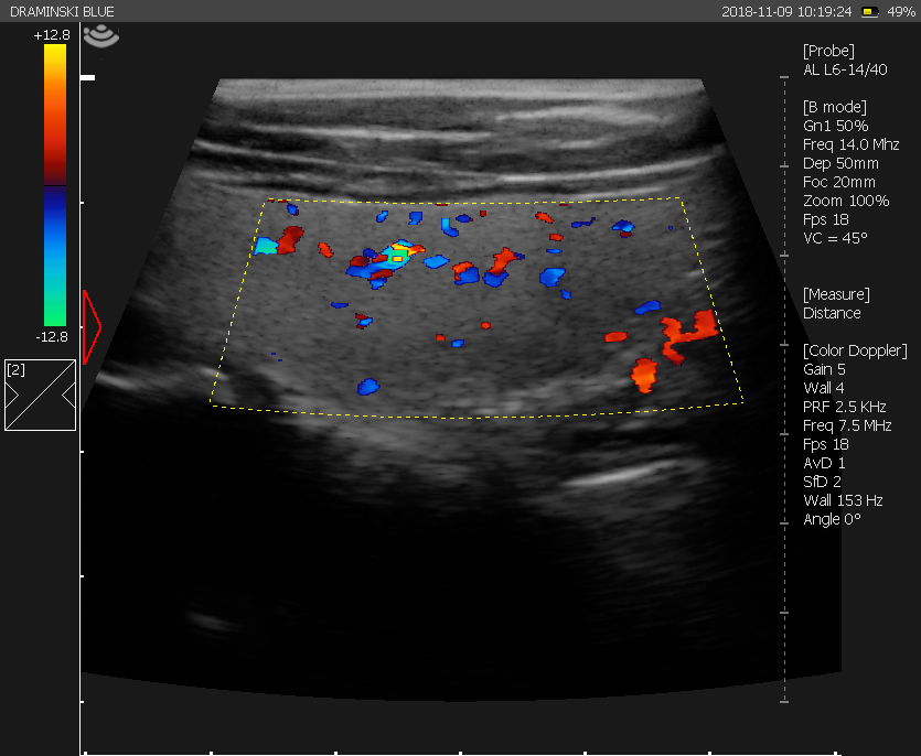
Blood flow in right lobe of thyroid gland
|
 |
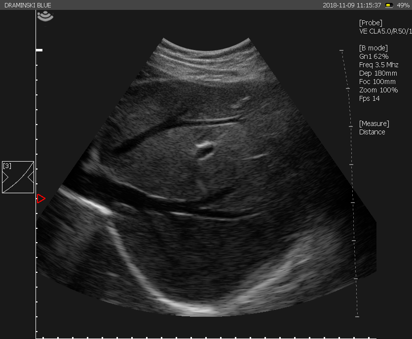
Hepatic veins |
 |
Case study #1 |
| A patient with myelodysplastic syndrome and symptoms of increasing dyspnoea, low fever and cough. During LUS there were visible an atelectatic and inflammatory subpleural consolidations with coexisting B lines in the course of interstitial pneumonia. |
Case study #2 |
| A 30-year-old patient was hospitalized due to increasing shortness of breath, weight loss, and progressive weakness. Mediastinal tumor in the course of DLBCL lymphoma. Visible solid change, hypoechogenic with mixed echostructure. Anechoic parts may correspond to areas of necrosis within the lesion. A visible piece of the left ventricular muscle adjacent to the lesion. |
Case study #3 |
| A patient with dilated cardiomyopathy – a severe left ventricular systolic dysfunction, hyperechoic thrombotic material visible within the apex in the right ventricle lumen. An increased accumulation of free fluid in the pericardial sac. Visible thrombus near the apex in the lumen of the right ventricle, also with the use of a linear probe. |
Case study #4 |
| A patient with chronic heart failure. Shortness of breath increasing over the past week. Free fluid of about 700 ml in the pleural cavity resulting in non-atelectral consolidation with pressure in the area of the lower right lung. Normal vascularization within consolidation. |